anatomy-of-the-skin
-
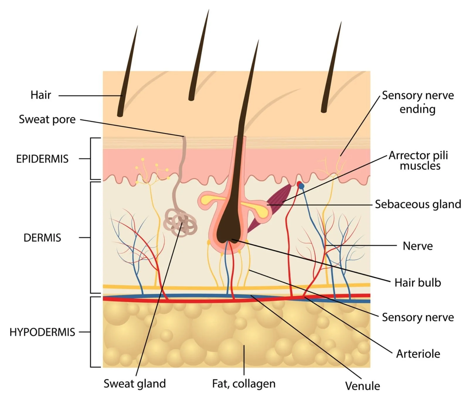
Dermis - The dermis also varies in thickness depending on the location of the skin. It is .30mm on eyelid and 3.0mm on the back, the dermal layers of the skins area complex tissue framework consisting of blood vessels, sweet glands, oils glands, nerves, hair follicles and where skin tissues like collagen and elastin are found. Collagen and elastin are proteins that help to maintain the skin's firmness and elasticity. The dermis is composed of three types of tissue (collagen, elastic, tissue and reticular).
Specialized Dermal Cells - The dermis contains many specialized cells and structures.
The hair follicles are situated here with the arrector pili muscle that attaches at each follicle.
Sebaceous (oil) glands and apocrine (scent) glands are associated with the follicle.
The layer also contains arrector (sweat) glands, but they are not associated with hair follicles.
Blood vessels and nerves course through this layer. The nerves transmit sensations of pain, itch, and temperature.
There are also specialized nerve cells called Meissner and VaterPacnini corpuscles that transmit the sensations of touch and pressure.
-
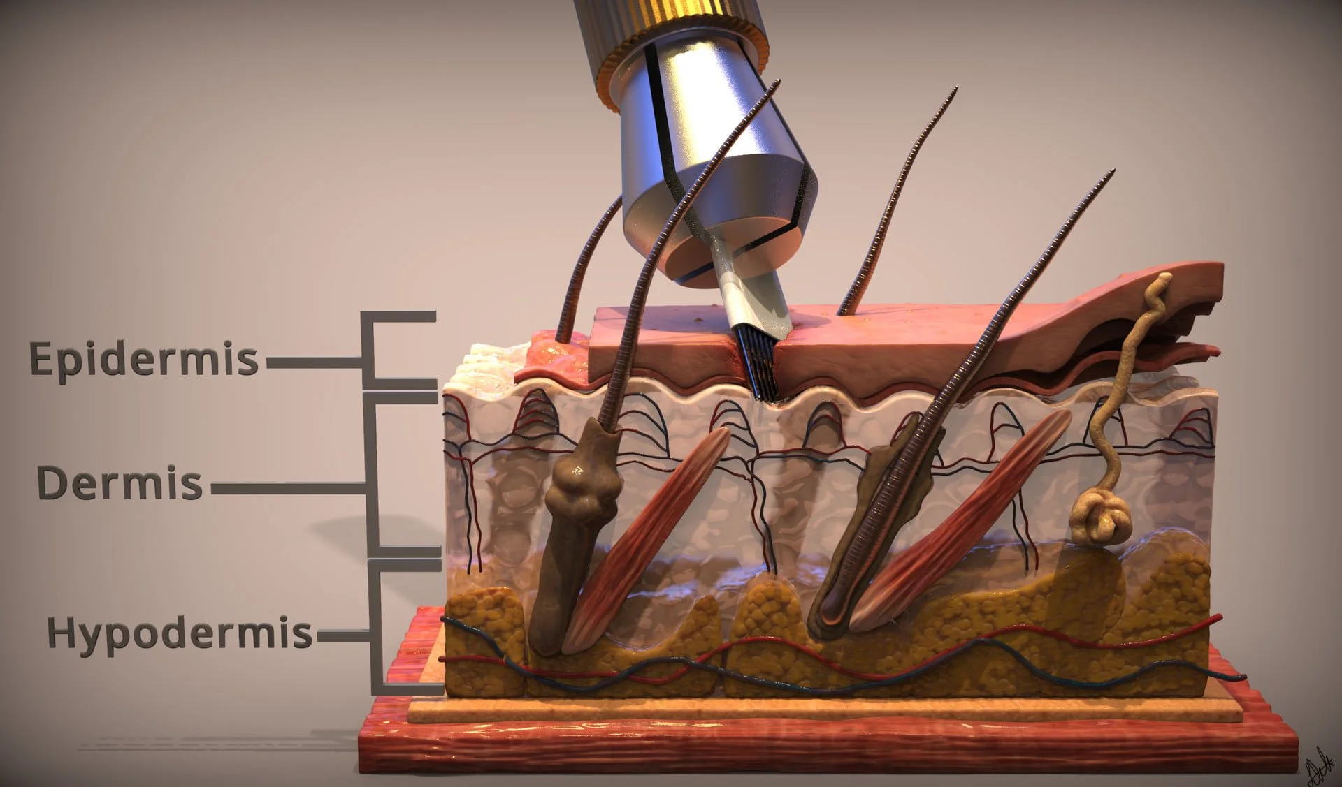
Traditional methods of permanent makeup consist of implanting pigment to the dermis. The dermis is live skin made up of blood vessels, sebaceous glands, sweat glands and nerves. Since this layer of skin is alive, it usually retains pigments permanently. In microblading, pigment is deposited right above the dermis into the basal layer of the epidermis. Because the cells of the basal layer continuously divide, the pigment is then broken down and eventually migrates to the surface causing the pigment to fade away.
The epidermis (the uppermost layer of skin) is an important system that creates our skin tone, while the dermis (the middle layer) contains connective tissue, hair follicles, and sweat glands that help regulate the integrity and temperature of the skin. The deeper hypodermis is made up of fat and even more connective tissue. The top five layers of the skin are called:
Basale
Spinosum
Granulosum
Lucidum
Corneum
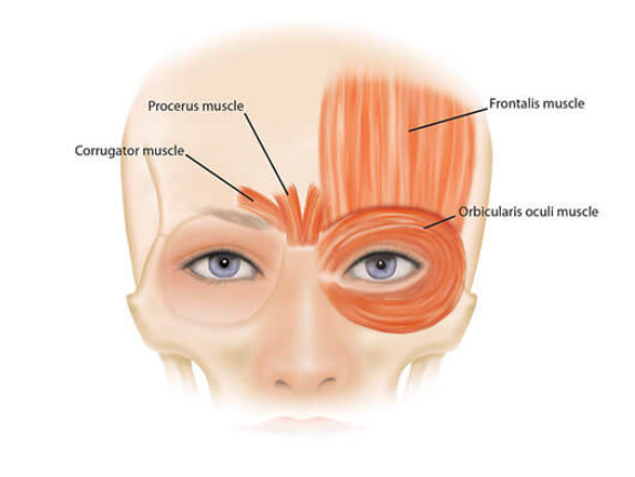
eyebrow Muscle Anatomy
The anatomical term for eyebrows is supercilia (singular: supercilium) from Latin. Superciliaalso refer to plumage around the eyes of certain birds!
Orbicularis Oculi
Is a muscle in the face that closes the eyelids. It arises from the nasal part of the frontal bone, from the frontal process of the maxilla in front of the lacrimal groove, and from the anterior surface and borders of a short fibrous band, the medial palpebral ligament.
Corrugator Supercilii
Is a small, pyramidal muscle that belongs to the circumorbital and palpebral group of facial muscles, along with the levator palpebrae superioris and orbicularis oculi muscles. Like other facial muscles, corrugator supercilii is innervated by branches of the facial nerve .
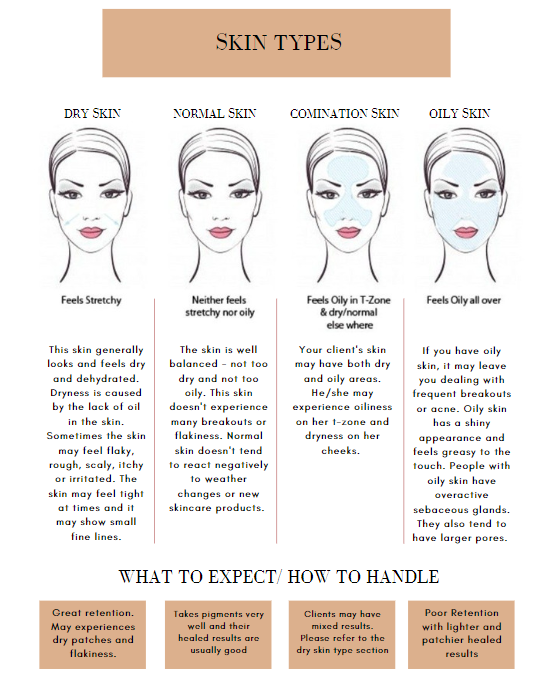
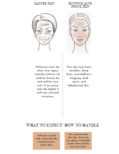
SKIN TYPES